FOOT MELANOMA - SIGNS TO WATCH FOR
posted: Aug. 01, 2018.
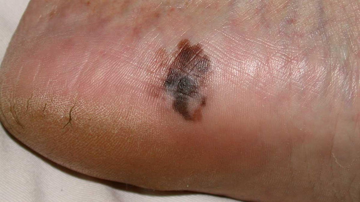
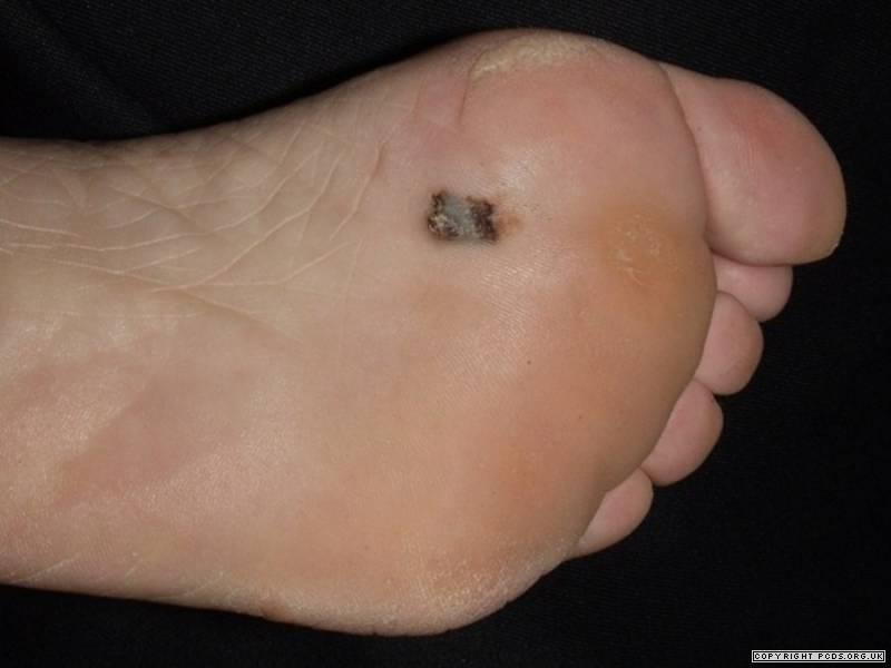
The development of new or changing pigmented lesions is the classic initial presentation of melanoma. A lesion that demonstrates a noticeable increase in size over a period of weeks to months might be suspicious. If the lesion is also changing in pigmentation (black, brown, red, blue or white) this also can be considered suspicious and should be evaluated by a practitioner - such as a chiropodist, dermatologist or physician and it should also be biopsied.
Foot melanoma is a type of skin cancer that affects the feet. It can appear anywhere on the foot, including the sole or under a nail. It starts in a type of skin cell called a melanocyte.
These cells are located in the uppermost layer of the skin. They are responsible for producing melanin, a dark pigment that helps screen the body against the harmful effects of ultraviolet light.
Foot melanoma is often treatable in the early stages, but it is often diagnosed late because people do not notice it. If it spreads, it may be life-threatening.
There are four different types of melanoma of the foot, these include: Acral lentiginous melanoma, Superficial spreading melanoma, Nodular melanoma and Amelanotic melanoma.
If you or a loved one has a suspicious mole/lesion on their foot it is important to get it assessed by a chiropodist and to also watch it cautiously for any signs of change and symptoms.
Brought to you by Doctor John A. Hardy, owner of Toronto's foot clinic, Academy Foot and Orthotics Clinic.